






-
Immunology(Colloidal Gold)Qualitative Test Cassette
Testing for Respiratory Diseases Testing for Infectious Diseases Testing for Tropical Infectious Diseases Testing for Tumor Markers Testing for Sexually Transmitted Diseases(STD) Testing for Pregnancy Testing for Drug Abuse Other Colloidal Gold Test Item List
-
Immunology(immunofluorescence chromatography)Quantitative Test Cassette
Testing for Myocardial Markers Testing for Reproductive Hormones Testing for Blood Glucose Testing for Vitamins Testing for Renal Function Other Immunofluorescence Test Item List
-
Immunofluorescence,Morphological chromogenic reagent
Pathogenic microorganism fluorescent staining solution Immunofluorescence staining reagent
-
Tissue and Cytology Staining Solution
Bacterial staining reagents Cell Tissue Staining Reagents Special staining reagent
-
Test Equipment and Scientific Research Instruments
Fluorescence Morphology Intelligent Inspection Platform Immunofluorescence POCT Analyzer Hematological Immun Analyzer Biochemical Immun Analyzer Chemiluminescence POCT Immun Analyzer Microbial Culture and Analysis System Microscopic Optics Pet Detection
-
Medical Consumables
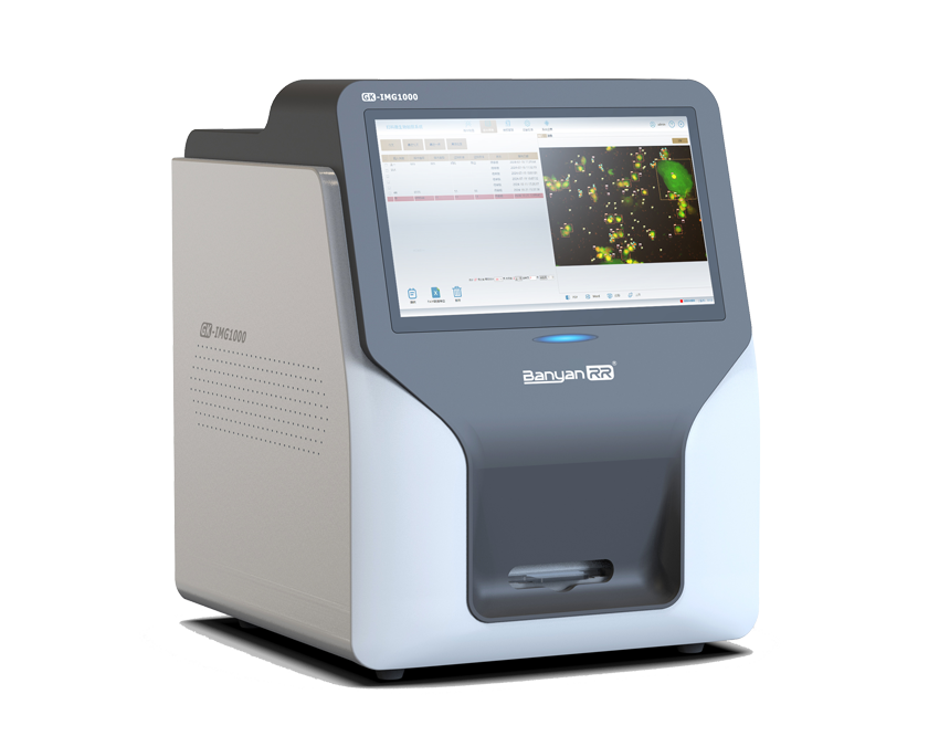
Product Name: Fluorescence Microscopy Image Scanning Analyzer
Model Numbers: GK-IMG1000, GK-IMG3000, GK-IMG5000, GK-IMG1000A, GK-IMG3000A, GK-IMG5000A
Detection Principle:
This device employs a detection method combining fluorescence microscopy with image scanning. After samples are stained with fluorescent dyes, cells within the sample emit specific fluorescent signals when excited by a fluorescent light source. The microscope optical system and image scanning system capture these signals to generate magnified, high-resolution fluorescent images. Utilizing convolutional neural network-based AI algorithms, the system processes scanned images, extracts relevant signals, and accurately identifies sample morphology, fluorescence intensity, and distribution characteristics. Through big data computation, quantitative analysis is performed, eliminating human observation errors and ensuring precise, stable results. This enables physicians to clearly observe pathogenic microorganisms such as fungi, trichomonads, clue cells, and Mycobacterium tuberculosis in samples, providing valuable support for clinical diagnosis.
Product Application:
This instrument enables fluorescent morphological detection of three types of pathogenic microorganisms and features an integrated touchscreen system.
1. Fungal Detection
The device can perform fully automated, standardized testing on various clinical samples such as human secretions, skin scales, nail debris, and sputum. By automatically staining specific fungal hyphae and spore core structures, combined with full-field automatic scanning and AI-based intelligent recognition technology, it accurately distinguishes fungal targets from sample impurities and dust interference. It rapidly determines the presence of fungal infection and quantifies positive detection results. Primarily used in hospital dermatology and laboratory departments, it supports screening and clinical diagnosis for superficial and deep fungal infections, as well as fungal skin diseases. The system effectively replaces traditional manual microscopy, significantly reducing the risks of missed or false detections while enhancing standardization and accuracy in fungal testing.
II. Vaginal Secretion Testing
The device is designed for fully automated fluorescent microscopic screening and quantitative analysis of gynecological vaginal secretion samples. Leveraging fully automated sample processing, it automatically handles sample loading, slide preparation, staining, and fluorescence scanning. By combining fluorescent labeling with AI-powered intelligent recognition algorithms, the system accurately identifies key targets in the sample—including fungi, trichomonads, white blood cells, epithelial cells, rod-shaped bacteria, and other microorganisms—automatically performing target detection, classification, counting, and intelligent interpretation. It enables efficient routine screening for gynecological vaginitis and reproductive tract infections, precisely assessing the vaginal microecological environment, microbial imbalances, and infection types. By eliminating human operational errors and subjective interpretation biases, it achieves standardized, intelligent, and high-throughput clinical testing for gynecological secretions.
III. Mycobacterium Tuberculosis Detection
Suitable for detecting clinical tuberculosis samples such as sputum and bronchoalveolar lavage fluid, the device performs fully automated staining and panoramic scanning imaging. Using specific fluorescent staining, Mycobacterium tuberculosis exhibits characteristic fluorescent morphology, which, combined with high-magnification panoramic scanning technology and AI-based intelligent recognition algorithms, allows precise capture of the bacterium’s distinctive cellular features. The system automatically identifies positive fields, counts bacterial numbers, and completes tuberculosis sample screening and accurate positive determination. Widely used in hospital initial tuberculosis screening, large-scale community screening, clinical diagnostic support, and treatment efficacy evaluation, this method offers full-field coverage, higher sensitivity, and more consistent results compared to traditional manual acid-fast staining microscopy. It is well-suited for routine, high-throughput tuberculosis testing in clinical settings.